| Biomedical Hypertexts Home | Glossary |
|---|
 |
Aquaporins: Water Channels |
|
Water crosses cell membranes by two routes: by diffusion through the lipid bilayer and through water channels called aquaporins. Functional characterization of the first aquaporin was reported in 1992, but water channels were suspected to exist well before that time, because the osmotic permeability of some types of epithelial cells was much too large to be accounted for by simple diffusion through the plasma membrane. A single human aquaporin-1 channel facilitates water transport at a rate of roughly 3 billion water molecules per second. Such transport appears to be bidirectional, in accordance with the prevailing osmotic gradient. The classical aquaporins transport solute-free water across cell membranes; they appear to be exclusive water channels and do not permeate membranes to ions or other small molecules. Some aquaporins - known as aquaglyceroporins - transport water plus glycerol and a few other small molecules. The Aquaporin Family
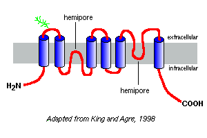
More than 10 different mammalian aquaporins have been identified to date, and additional members are suspected to exist. Closely related water channel proteins have been isolated from plants, insects and bacteria. Aquaporin-1 from human red blood cells was the first to be discovered and is probably the best studied. Based on hydrophobicity plots of their amino acid sequences , the aquaporins are predicted to have six membrane-spanning segments, as depicted in the model of aquaporin-1 to the right. Based on studies with aquaporin-1, it appears that aquaporins exist in the plasma membrane as homotetramers. Each aquaporin monomer contains two hemi-pores, which fold together to form a water channel. Different aquaporins have different patterns of glycosylation. In the case of aquaporin-1, the peptide backbone is roughly 28 kDa and the glycosylated forms range from 40 to 60 kDa in mass. Most aquaporins have a protein kinase A phosphorylation motif in one of the cytoplasmic loops, and differential phosphorylation is suspected empart a regulatory function to the molecule. Patterns of Aquaporin ExpressionEach of the aquaporins has an essentially unique pattern of expression among tissues and during development. A summary of these attributes and some of the important potential or known functions is presented in the following table: |
| Major Sites of Expression | Comments | |
|---|---|---|
| Aquaporin-0 | Eye: lens fiber cells | Fluid balance within the lens |
| Aquaporin-1 | Red blood cells | Osmotic protection |
| Kidney: proximal tubule | Concentration of urine | |
| Eye: ciliary epithelium | Production of aqueous humor | |
| Brain: choriod plexus | Production of cerebrospinal fluid | |
| Lung: alveolar epithelial cells | Alveolar hydration state | |
| Aquaporin-2 | Kidney: collecting ducts | Mediates antidiuretic hormone activity |
| Aquaporin-3 * | Kidney: collecting ducts | Reabsorbtion of water into blood |
| Trachea: epithelial cells | Secretion of water into trachea | |
| Aquaporin-4 | Kidney: collecting ducts | Reabsorbtion of water |
| Brain: ependymal cells | CSF fluid balance | |
| Brain: hypothalamus | Osmosensing function? | |
| Lung: bronchial epithelium | Bronchial fluid secretion | |
| Aquaporin-5 | Salivary glands | Production of saliva |
| Lacrimal glands | Production of tears | |
| Aquaporin-6 | Kidney | Very low water permeability; function? |
| Aquaporin-7 * | Testis and sperm | |
| Aquaporin-8 | Testis, pancreas, liver, others | |
| Aquaporin-9 * | Leukocytes | |
| * an aquaglyceroporin | ||
|
It should be clear that aquaporins are very widely distributed, and also that the different aquaporins have different functionally-important specialties. Several interesting and important features of aquaporin-mediated water transport are illustrated in the principal cells that line collecting ducts in the kidney. Water flowing through these ducts can either continue on and be voided in urine or be reabsorbed across the epithelium and back into blood. Reabsorption is essentially nil unless the epithelial cells see antidiuretic hormone, which strongly stimulates reabsorption of water. Collecting duct cells express at least two aquaporins:
 Aquaporins and DiseaseConsidering the importance of water transport in a myriad of physiologic processes, it is to be expected that lesions in aquaporin genes or acquired dysfunction in aquaporins may cause or contribute to several disease states. The search for such connections is still early, but two clear examples of disease have been identified as resulting from deficiency in aquaporins:
A small number of people have been identified with severe or total deficiency in aquaporin-1. Interestingly, they appear clinically unaffected, but have not been examined under conditions of water deprivation. Mice with targeted deletions in aquaporin-1 also appear normal and healthy unless they are fluid restricted, in which case they become severely hyperosmolar. |
 |
References and Reviews
|
Last updated on Jan 2, 2002 |
| Author: R. Bowen |
| Send comments via form or email to rbowen@lamar.colostate.edu |