PLANT ANATOMY LAB III
EXERCISE SHEET
HAND SECTIONING AND HISTOLOGY OF CELL
CONTENTS
Unlock
c:ANATOMY/Ch2_Ultrastructure/lab3exercise.html
Use SeaMonkey Composer to complete the
exercise sheet - Click on File, then Edit Page.
Make sure you save your changes by
replacing
the existing file, (Click on File, then Save) else all
will be lost!
NAME _____________________________.
(107 points)
I. Elodea leaf
-
A. Immediately after mounting my
leaf
specimen the distribution of
chloroplasts in the leaf cells was __________ (1 point)
-
But
after ___
minutes they ________ (2 points)
-
I
think that this occurred because ______ (1 point)
-
B. I observed cytoplasmic
streaming
to occur first in ________ (1 point)
-
C. I was able to
distinquish chloroplasts
from nuclei since ______ (1 point)
-
D. I estimate the average cell
dimensions
expressed in ____ units in this leaf to be as follows: (1 point)
+
(9 points)
| PLANE OF ORIENTATION |
LENGTH OF CELL |
LENGTH OF CELL |
LENGTH OF CELL |
MEAN
LENGTH OF CELL |
| LONGITUDINAL |
|
|
|
|
| TRANSVERSE |
|
|
|
|
| THICKNESS |
|
|
|
|
-
E. Here are my best examples of
unstained
and stained Elodea leaf cells:
|
UNSTAINED
|
STAINED
|
I STAINED WITH
|
| (6 points) |
(6 points) |
|
| (6 points) |
(6 points) |
|
- F. I
have used one of the above images with Image-J to re-estimate the
average cell dimensions in ____
units in this leaf to be as follows: (1 point) +
(6 points)
| PLANE OF ORIENTATION |
LENGTH OF CELL (Image-J)
|
LENGTH OF CELL (Image-J) |
LENGTH OF CELL (Image-J) |
MEAN
LENGTH OF CELL (Image-J) |
| LONGITUDINAL |
|
|
|
|
| TRANSVERSE |
|
|
|
|
-
G. Some of the benefits in using
general
histological stains include ________ (2 points)
-
H. Some of the disadvantages in
using
general histological stains include _________ (2 points)
-
I. I can tell a cell is
living
because _____________ (2 points)
II.
Cell division.
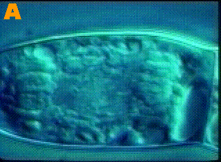
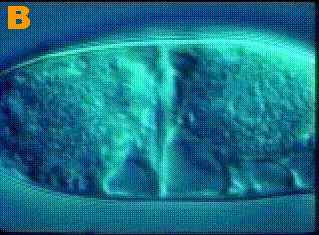
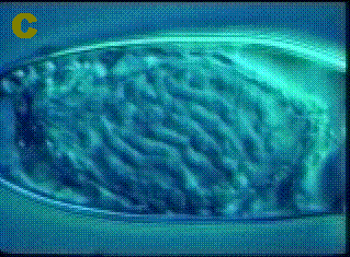
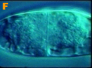
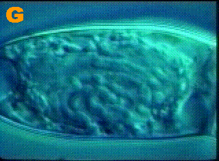
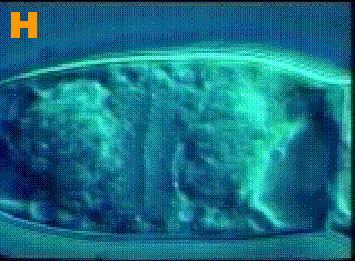
The sequence of these images of a dividing
cell are out of order!
Right click on one of the images
above,
then cut, move to the appropriate cell in the table below, then right
click
and paste the image where you think it belongs in the sequence.
Correct Temporal Sequence of Cell
Division:
(6 points)
| First |
Second |
Third |
| Fourth |
Fifth |
Sixth |
^
Moving from left to right, this is the
sequence in which cell division really occurs! I've even
labeled
the stages!
III and IV. Below are my best
examples
of the indicated cellular structures:
| SUBJECT |
IMAGE |
SUBJECT |
IMAGE |
Chloroplast (___X)
from _______ |
(6 points) |
Chromoplast (___X)
from _______ |
(6 points) |
Leucoplast (___X)
from _______ |
(6 points) |
_____________Crystal (___X)
from _______ |
(6 points) |
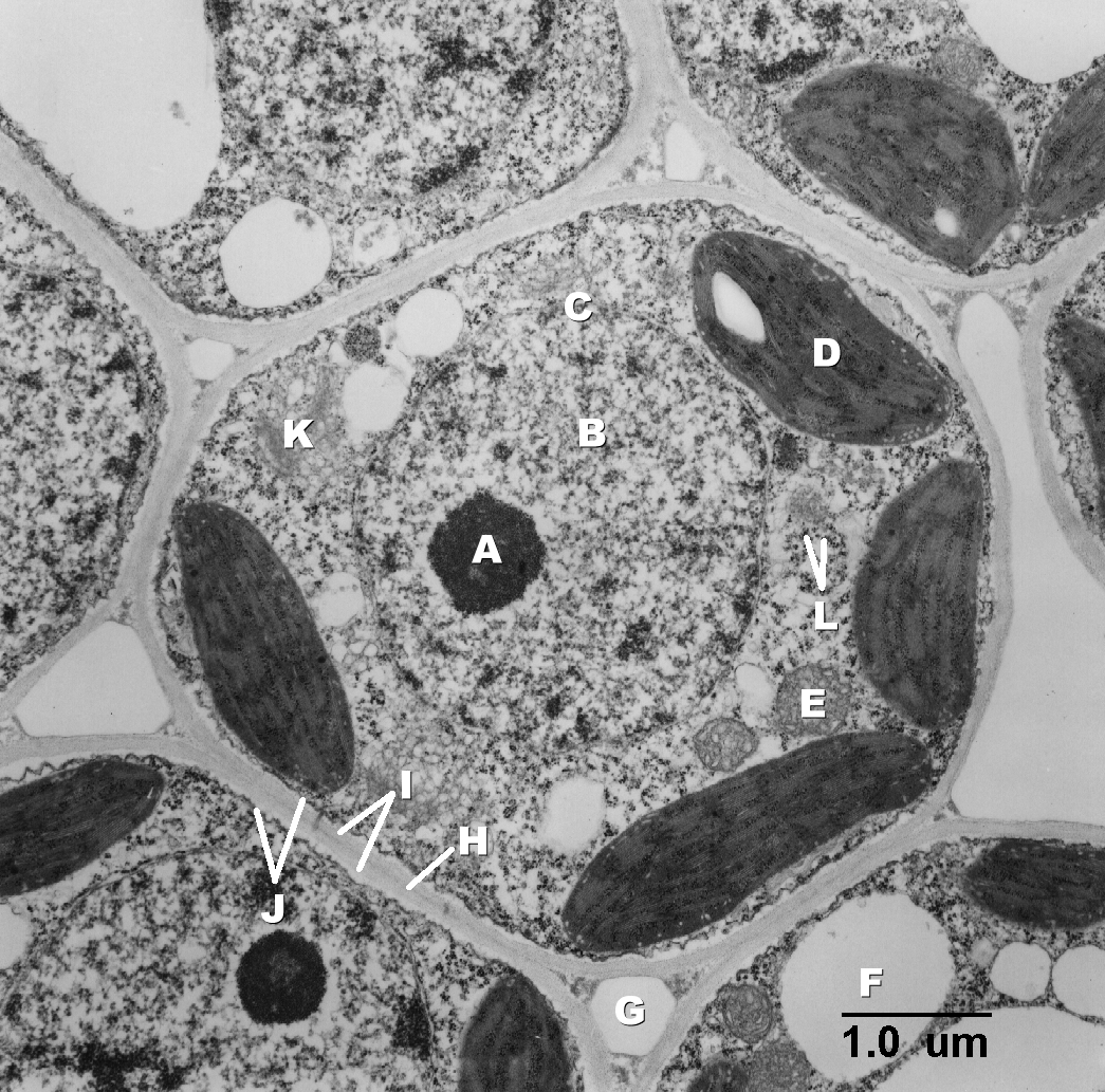
V. I have labeled the indicated
cellular
structures in the transmission electron
micrograph
of
a typical plant cell and provided my best example of a bright field
light
micrograph of a plant cell on which I have labeled those cellular
structures
that are resolvable with the compound light microscope: (12
points) + (12 points)
|
RESOLVABLE WITH T.E.M.
|
NAMES OF INDICATED STRUCTURES
|
MY IMAGE OF A PLANT CELL ON
WHICH I HAVE
LABELED STRUCTURES THAT ARE RESOLVABLE WITH LIGHT MICROSCOPY
|
 |
| A. |
| B. |
| C. |
| D. |
| E. |
| F. |
| G. |
| H. |
| I. |
| J. |
| K. |
| L. |
|
|
