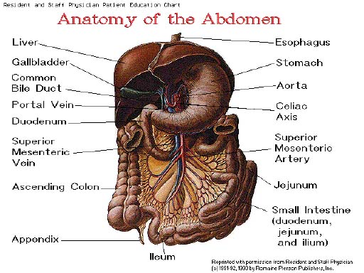

Digestive Processes
food prepared for consumption by the cells through 5 basic
activities
1. Ingestion--taking food
into the body
2. Movement--passage of
food along the gastrointestinal tract
3. Digestion--breakdown of
food by both mechanical and chemical processes
4. Absorption--passage of
food from the GI tract into the cardiovascular and lymphatic
systems for distribution to the cells who need nutrients
5. Defecation--elimination
of indigestible substances from the GI tract
Chemical digestion
series of catabolic reactions that break down large molecules
(carbohydrates, proteins, lipids) into smaller molecules that can be
absorbed and used by the body
Mechanical digestion
-movements of the GI tract that aid in overall digestion
-teeth prepare food to be swallowed and smooth muscle of the stomach
and small intestine churn the food so it mixes with enzymes that help
in the chemical reaction
1. Gastrointestinal
Tract (GI tract)
-also referred to as the alimentary canal
-continuous tube running through the ventral cavity from the mouth to
the anus
-average length is 9 meters
-organs include: mouth, pharynx, esophagus, stomach, small intestine,
large intestine
2. Accessory Structures
-teeth, tongue, salivary glands, liver, gall bladder, pancreas (all
accessory structures lie outside the GI tract with the exception of
the teeth and tongue)
-these structures produce or store secretions that aid in the
chemical breakdown of food
-secretions reach the GI tract through ducts
4
basic layers throughout the
GI tract from the esophagus to the anus
1. Mucosa--inner lining of the
GI tract, mucous membrane
2. Submucosa
-dense connective tissue that binds the mucosa to the muscularis
-highly vascular and contains part of the autonomic nerve supply to
the muscularis mucosa
3. Muscularis
-this layer in the mouth, pharynx and upper esophagus consist of some
skeletal muscle that produces voluntary swallowing
-rest of the GI tract consist of smooth muscle found in 2 sheets
inner sheet contains circular fibers outer sheet contains
longitudinal fibers
-helps to fix food and move it through the GI tract
4. Serosa
-outermost layer of the GI tract
-also called peritoneum
one extension of the parietal peritoneum is called the

mouth (oral cavity, buccal
cavity)
formed by the checks, hard palate, soft palate and tongue
uvula-- tissue that protrudes
past the end of the soft palate
tongue--
-accessory structure
-composed of skeletal muscle covered by mucous membranes
Mastication (mechanical
process)
-tongue moves food, teeth grind food, food mixes with saliva
-food reduced to a soft flexible bass called a
Bolus
Salivary amylase
-starts the breakdown of starch (only chemical digestion that occurs
in the mouth)
3 stages
1. voluntary
-bolus forced to the back of the mouth cavity and into the oropharynx
by the tongue pressing up and back against the palates
2. pharyngeal
-bolus stimulates nerves in the oropharynx
-impulses cause soft palate and uvula to move up and seal off the
nasopharynx
3. esophageal
-bolus pushed through the esophagus by involuntary muscular movements
called peristalsis

*stomach can only absorb some water, electrolytes, certain drugs
(aspirin) and alcohol
histology
made of the same four layers as the rest of the GI tract (these
layers are modified)
rugae--large folds of an empty
stomach
chief cells--secrete pepsin,
which is as enzyme
parietal cells--secrete HCL
and intrinsic factor-- helps
absorption of vitamin B12
mucous cells
enteroendocrine cells
-produce gastrin--
**collection of above juices is called gastric juice
-mixing waves (peristalic movements) pass over the stomach every
15 to 25 seconds
-gastric juices reduce bolus to a thin liquid called
chyme
-excess food stored in the fundus
-mixing waves force chyme toward the pyloric sphincter
-sphincter only allows a portion of the chyme to pass through
Chemical digestion
-starts the breakdown of protein
-pepsin breaks the peptide bonds that hold the amino acids
together
-pepsin only works in an acidic environment (stomach pH of 2)
-alkaline mucous lines the stomach which should prevent the breakdown
of the stomach walls
3 phases trigger gastric secretions
1. cephalic (reflex)
phase--
-before food enters the stomach
-sight, smell, taste or thought of food stimulates production of
gastric juices
2. gastric phase--
-food enters the stomach
-distention of the stomach triggers this phase
-partially digested proteins and caffeine stimulate secretion of
3. intestinal phase--
-chyme enters the intestine 2-6 hours after ingestion
-overall effect is the inhibition of gastric secretions
-three hormonal secretions cause the inhibition of gastric juices
pancreas
-accessory structure of the GI tract
-oblong gland 12.5 cm long and 2.5 cm thick
-lies posterior to the greater curvature of the stomach
-connected by 2 ducts to the upper part of the small intestine
liver (LD)
-about 1.4 Kg in an average adult
-under diaphragm in the right hypochondrium and part of the
epigastrium of the abdomen
-2 principal lobes
histology
lobules--
-functional unit of the liver
-each lobule surrounds a central vein
-cells of the lobule secrete:
-mostly H2O with bile salts
-lining of spaces between cells are phagocytic
Kupffer’s cells--
-destroy worn out RBC and white blood cells, bacteria and toxic
substances
-filters blood (all things absorbed from the GI tract go to the liver
first)
physiology
1. carbohydrate
metabolism--maintains normal blood glucose levels
2. fat
metabolism---synthesizes cholesterol and digest cholesterol
and stores fat
3. protein metabolism--
-loss of this function results in death with in a few days
-removal of nitrates
-conversion of NH3 into urea
-synthesis of plasma proteins
-synthesis of fibrenogen (blood coagulant)
-synthesis of anticoagulants
-conversion of one amino acid to another
4. removal of drugs and
hormones
-detoxify penicillin, ampicillin
-excrete or alter into bile, estrogen, or aldosterene
5. excretion of bile
6. synthesis of bile salts
7. storage--glycogen, vitamins
A, B12, D, E, K, iron, copper
8.
phagocytosis--Kupffer’s
cells
9. activation of vitamin D
gallbladder
-pear shaped sac about 7-10 cm long
-accessory structure
-located in the fossa of the visceral surface of the liver
physiology
-hormonal stimulation causes smooth muscle contraction and squeezes
contents into the cystic duct
and common bile duct
-stores and concentrates bile until small intestine needs
-empty small intestine = closed hepatopancreatic ampulla
sphincter
(bile flows back to gallbladder)
-chyme in small intestine opens the sphincter
small intestine
-2.5 cm in diameter, 6.35 m long
-absorption and most digestion occur here
-intestinal glands--secrete
juices
-duodenal glands--secrete
alkaline mucous
large surface area = better
absorption
4 things help the small intestine absorb better
1. length
2. microvilli
3. villi--
-projections about 1 mm high
-10 to 40 every square mm
-gives lining of the small intestine a velvety appearance
-each contain: arteriole, venule, capillary network, and lymphatic
vessels
4. circular folds
(plicae circulares)
-permanent ridges about 10 mm high
-start near the proximal portion of duodenum and stop at the midpoint
of ileum
-causes chyme to “spiral” which enhances absorption by
increasing the contact with villi
mechanical
1. segmentation--
-major movement of small intestine
-localized contraction to mix the chyme
-DOES NOT move chyme downward
2. peristalsis--
-moves chyme down
-weak compared to in the esophagus
-chyme moves at 1 cm per minute (food stays in the small intestine
about 3 to 5 hours
chemical
-pancreatic juice, bile and intestinal juice complete the breakdown
of carbohydrates, proteins and lipids
-food pieces need to be small enough to pass through the
epithelial tissue of the villi and depends on diffusion, facilitated
diffusion, osmosis, and active transport
carbohydrates --- monosacharides
proteins --- amino acids
fats, lipids --- fatty acids, glycerol, and glycerides
Monosacharides and Amino acids
go into the blood -- liver -- then to general circulation.
Fatty acids and Glycerides--
-bile salts dissolve and form bubbles called
micelles--
-container for fatty acids and glycerides
-when they come in contact with epithelial cells they allow contents
to diffuse out of the “bubble”
-the micelle stays in the small intestine
Lipids hitch rides with
protein transporters to form lipoproteins
Water
-absorbed by osmosis
-on average, 9 liters of water enters the small intestine a day
-all but 1 liter is absorbed
Large
Intestine
-completes absorption (less than 10%)
-forms and expels feces
Anatomy
1.5 meters in length and 6.5 centimeters in diameter
Cecum
-portion below the ileocecal valve
-the appendix is attached to the pouch of the cecum
Colon--divided into 4
parts
Ascending -- up
Transverse -- across
Descending -- down
Sigmoid -- angles toward the midline of the body
Rectum--last 20 centimeters of
the gastrointestinal tract
Anal Canal--last 2 to 4
centimeters of the rectum
Anus
-opening of the anal canal
-internal anal sphincter -- smooth “involuntary muscle”
-external anal sphincter -- skeletal muscle
Histology
-same four layers as the rest of the GI tract
-no villi
-no circular folds
simple columnar epithelial cells absorb
water
goblet cells secrete mucous
Digestion
Chemical
-no enzymes (digestion through bacteria action)
-bacteria ferment excess carbohydrates and this releases hydrogen,
methane and carbon dioxide
-the bacteria breakdown of amino acids produce odor
Feces formation
-chyme stays in the large intestine for 3 to 10 hours, during which
it is a semi-solid due to the absorption of water
-all but about 100 milliliter of water is absorbed
Feces
-consist of water, inorganic salts, sloughed off epithelial cells,
products of bacteria decomposition and undigested parts of food
Defecation
-emptying of rectum
-triggered by distention of the rectum
|
|
|
|
|
|
|
|
|
|
|
|
|
|
|
|
|
|
|
|
|
|
|
|
|