Bacteria, like most cells, are essentially transparent, and must be stained
in order to be easily visualized under the microscope. The specimen must be
first spread out on a clean slide (preparation of a smear), heated gently to fix
it to the slide, and then stained with an appropriate dye. Bacteria tend to be
negatively charged, therefore positively-charged or basic dyes will bind to and
stain them.
EQUIPMENT AND SUPPLIES:
clean microscope slide
soap and hot water
dH2O
in dropper bottle
sample to be smeared
bacteriological
loop (26 gauge Platinum as specified in Equipment
for a Microbiological Work Station)
flame source
stains: 0.3%
methylene blue or
Hucker's Crystal Violet
tap water
paper
towels or bibulous paper
PREPARE
THE SMEAR: |
|
 |
1. Secure a CLEAN microscope slide. The smear will not spread out
properly if the slide is even slightly oily. If in doubt, wash slide well
with soap and water, polish
with clean paper towel or a Kimwipe (do not use brown recycled towels,
they give off too much lint). |
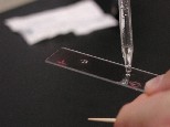 |
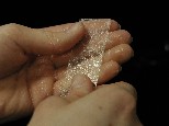
2. a. If the specimen is solid: place a small drop of
dH20 on a clean slide. Pick up small sample with a sterile loop
and suspend in the water. Spread to the diameter of a dime (1.8 cm). The
suspension should appear very faintly cloudy. |
|
2. b. If liquid: place small drop of sample on microscope
slide, spread to the diameter of a dime. Dilute with small drop of water
if more than faintly turbid. |
| FIX THE SMEAR: |
|
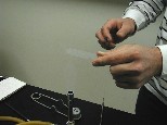 |
3. Fix smear by lightly passing slide through a gentle flame. Do not
overheat. The slide should not get too hot to comfortably hold. You
are just trying to dry out the sample so that it will stick to the slide
and not wash off in the next two steps. |
STAIN THE
FIXED SMEAR: |
|
 |
4. Add a drop or two of stain (for instance, 0.3% methylene blue or
Hucker's Crystal Violet). Let sit 1 minute (or 30 seconds for Hucker's
Stain, a more powerful stain). |
|
5. Gently rinse off excess stain with tap water for a few seconds
until no more stain is seen to flow off. Gently blot dry (do not rub) with
clean white paper towel. (Bibulous paper should be used if lint is a
problem.) |
VIEW THE
STAINED SMEAR: |
|
|
6. Examine with 10x objective to locate a region which is well spread
and not too heavily stained. View briefly with 40x objective to confirm a
well-spread, well-stained section. Then use 100x oil immersion objective
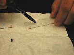
to study specimen (FOLLOW Oil Immersion Protocol CAREFULLY). Adjust
light for maximum clarity and carefully draw individual bacteria with all
structures seen. Title the drawing as to sample, give the stain used and,
at lower right, indicate the magnification of the illustrated view. |
|
7. If you are not saving your slide, wash it well in hot soapy water,
air dry in a plastic rack. |
Suggestions for specimens to view: Buccal smear, tooth scrapings, yogurt,
buttermilk, yeast, fecal smear, vaginal smear, single colonies from agar plates,
etc.
Go to Fankhauser's
Cheese Page
or
to David Fankhauser's Main
Page
Send Email to: FANKHADB@UC.EDU


